TL;DR
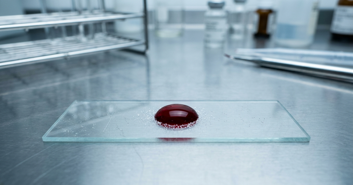
- The Study: Researchers at the University of Bristol showed in male rats that red blood cells (RBCs) exchange components of the endothelial glycocalyx — the fragile sugar-protein coating that lines blood vessels — and that an RBC blood draw reflects the state of that coating in heart and kidney microvessels.
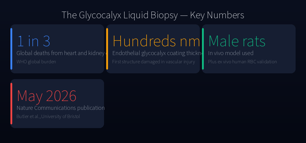
- Why It Matters: Heart and kidney diseases account for roughly one in three deaths worldwide, but today’s screening tools (eGFR, albuminuria, fasting glucose) usually flag damage long after microvessels are injured.
- The Caveat: This is a preclinical proof-of-concept in animals plus ex vivo human samples — not a clinical test. The actionable advice today is still blood pressure, glucose, lipids, salt and smoking.
The Vascular Lining Falls Apart First — and We Have No Easy Way to See It
For decades, cardiologists and nephrologists have known that the endothelial glycocalyx — the few-hundred-nanometer sugar-protein coating that lines the inside of every blood vessel — is one of the first structures to break down in vascular disease (Reitsma et al., Pflügers Archiv 2007). High blood sugar, high blood pressure, sepsis, smoking, oxidative stress: all of them shear or strip the glycocalyx before clinical biomarkers move.
The problem is measurement. Until now, three options existed, and all had limits:
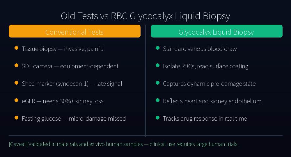
- Tissue biopsy — accurate but invasive, with real risks for patients.
- Sidestream dark-field (SDF) imaging of the sublingual microcirculation — equipment-heavy and operator-dependent.
- Shed-marker blood tests (syndecan-1, heparan sulfate fragments) — these only detect glycocalyx that has already been shed into the bloodstream. By that point, you are counting the rubble after the wall has come down.
That is the gap a paper published on May 12, 2026 in Nature Communications (Butler MJ et al., DOI: 10.1038/s41467-026-71848-4) tries to fill.
What the New Study Actually Did
Methodology Transparency
The Bristol team used azide-alkyne cycloaddition (click chemistry) to tag sugar residues on the glycocalyx with a fluorescent label that does not disturb the underlying biology. They tracked these tagged sugars across three preparations:
- In vivo, in male rats, observing capillary blood flow under intravital microscopy.
- Ex vivo, by drawing blood and isolating RBCs to measure glycocalyx components on their surface.
- In parallel, by directly assaying glycocalyx integrity and endothelial barrier permeability in the heart and kidney tissues of the same animals.
This three-arm design is what makes the study persuasive: the team did not just claim RBCs carry the glycocalyx signal — they showed that the RBC readout agreed with the gold-standard tissue measurements in the same animals.
Mechanism: A → B → C
The full pathway, end to end:
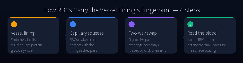
- A. Direct contact: As an RBC squeezes through a capillary that is roughly its own diameter, its surface and the endothelial lining make sustained physical contact.
- B. Bidirectional exchange: Click-labeled glycocalyx sugars moved from endothelial cells onto RBCs — and from RBCs onto endothelial cells. This is not passive shedding. It is a reciprocal swap that continues throughout circulation.
- C. Biochemical imprint: Because RBCs see every microvessel in the body, their surface accumulates a running average of glycocalyx state across heart, kidney and other beds. Damage in the endothelial coat shows up as altered RBC glycocalyx composition.
This matters because shed-marker tests (syndecan-1, etc.) sit at the end of the chain — they catch material only after it has been cleaved off. The RBC readout sits in the middle of the chain, capturing the dynamic equilibrium before the wall is fully damaged.
Competing Hypotheses — What This Reframes
The textbook model of vascular damage detection has been linear: insult (e.g., hyperglycemia) → glycocalyx loss → endothelial dysfunction → shed-marker rise → eGFR/albuminuria abnormality → clinical event. Most early-detection efforts target the later steps in that chain.
This work proposes a different point of measurement: the equilibrium between endothelium and RBC, which appears to track glycocalyx state continuously. If validated in humans, it would let clinicians sit closer to the first insult — not at the last visible step.
What the Study Did Not Do
Equally important: what the study did not establish.
- It did not test humans in vivo. The ex vivo human RBC data are supportive but limited.
- It used male rats only. Sex differences in endothelial biology are well documented and remain to be addressed.
- It did not demonstrate that RBC glycocalyx changes predict clinical events (heart attack, dialysis, death) over years of follow-up. That requires prospective human cohorts.
- The assay itself still depends on click chemistry and specialized labeling, which is not yet a routine clinical workflow.
These limits are not unusual for a first-in-kind methodological paper. They are the reason this is a paradigm-opening result, not a clinical announcement.
Old vs. New: How the Logic of the Test Changes
The proposed pipeline is simple: standard venous blood draw → isolate RBCs → quantify glycocalyx composition on the cell surface → estimate microvascular endothelial damage. Compared to current tools:
- Burden: A routine blood draw versus a biopsy or imaging session.
- Timing: Captures the active state of the glycocalyx, not the rubble after collapse.
- Coverage: RBCs traverse every microvascular bed, so the readout integrates heart, kidney and other tissues simultaneously — a notable advantage given how often heart and kidney disease move together.
- Drug monitoring: SGLT2 inhibitors, statins and sulodexide have all been associated with glycocalyx recovery (Targosz-Korecka et al., Scientific Reports 2020; Cherney et al., Circulation 2014). A faster RBC-based readout could let clinicians track whether a drug is actually restoring the lining in a specific patient.
What This Means For You
Until validated human assays arrive — and that is unlikely to be within the next two years — none of this changes clinical decision-making. What it does do is reinforce the mechanistic case for a set of habits we already know matter for the endothelium. With dose, timing and mechanism made explicit:
- Tighten glucose control to HbA1c < 7% if you have diabetes. Hyperglycemia activates neuraminidase that strips sialic acid from the glycocalyx (Nieuwdorp et al., Diabetes 2006). The UKPDS trial showed a roughly 25% reduction in microvascular complications with intensive glucose control (Lancet 1998).
- Target blood pressure < 130/80 mmHg if your physician agrees. Shear stress at high pressure mechanically degrades the glycocalyx; SPRINT (NEJM 2015) showed a 25% reduction in major cardiovascular events with more aggressive control.
- Stop smoking. Cigarette smoke produces oxidative stress that degrades the glycocalyx in hours. Endothelial function measurably improves within a year of cessation (Celermajer et al., Circulation 1996).
- 150+ minutes per week of moderate aerobic exercise. Pulsatile shear stress during exercise is one of the strongest known stimulants of glycocalyx synthesis (Mulivor & Lipowsky, Am J Physiol 2004).
- Reduce sodium toward ~2,000 mg/day. High sodium has been shown to degrade the endothelial glycocalyx independently of blood pressure in animal models (Olde Engberink et al., Hypertension 2018).
- If you have type 2 diabetes, heart failure or CKD, ask about SGLT2 inhibitors. Their glycocalyx-restorative signal in mechanistic studies is consistent with their clinical outcomes (Cherney et al., 2014).
The single most useful thing a non-patient can do right now is not skip the basics on your next physical: blood pressure, fasting glucose or HbA1c, lipid panel, eGFR, and urine albumin-to-creatinine ratio. None of these is as sensitive as the RBC glycocalyx assay might one day be — but they are what we have, and they catch many problems early enough to act.
The Bottom Line
The Bristol paper does not deliver a new blood test you can ask for tomorrow. It delivers something arguably more interesting: a different point on the timeline at which we can read vascular damage. The conceptual shift — from counting rubble to reading the equilibrium — is the kind of step that, if it holds up in humans, could change how cardiology and nephrology screen for risk over the next decade.
Expect to see this work followed by larger animal studies, human pilot cohorts, and eventually a standardized assay. Until then, the playbook for protecting the lining of your blood vessels is unchanged — and quietly better supported than it was a month ago.
This content is for informational purposes only and is not a substitute for professional medical advice, diagnosis, or treatment. Always seek the advice of your physician or other qualified health provider with any questions you may have regarding a medical condition.
References
- Butler MJ et al. Endothelial-erythrocyte glycocalyx exchange enables liquid biopsies of endothelial function. Nature Communications 2026; vol. 17, article 71848. DOI: 10.1038/s41467-026-71848-4.
- Reitsma S et al. The endothelial glycocalyx: composition, functions, and visualization. Pflügers Archiv 2007;454(3):345-359.
- Nieuwdorp M et al. Loss of endothelial glycocalyx during acute hyperglycemia. Diabetes 2006;55(2):480-486.
- Olde Engberink RH et al. The role of sodium in the endothelial glycocalyx. Hypertension 2018;71(6):986-994.
- Cherney DZ et al. Renal hemodynamic effect of SGLT2 inhibition in patients with type 1 diabetes mellitus. Circulation 2014;129(5):587-597.
- SPRINT Research Group. A randomized trial of intensive versus standard blood-pressure control. NEJM 2015;373:2103-2116.